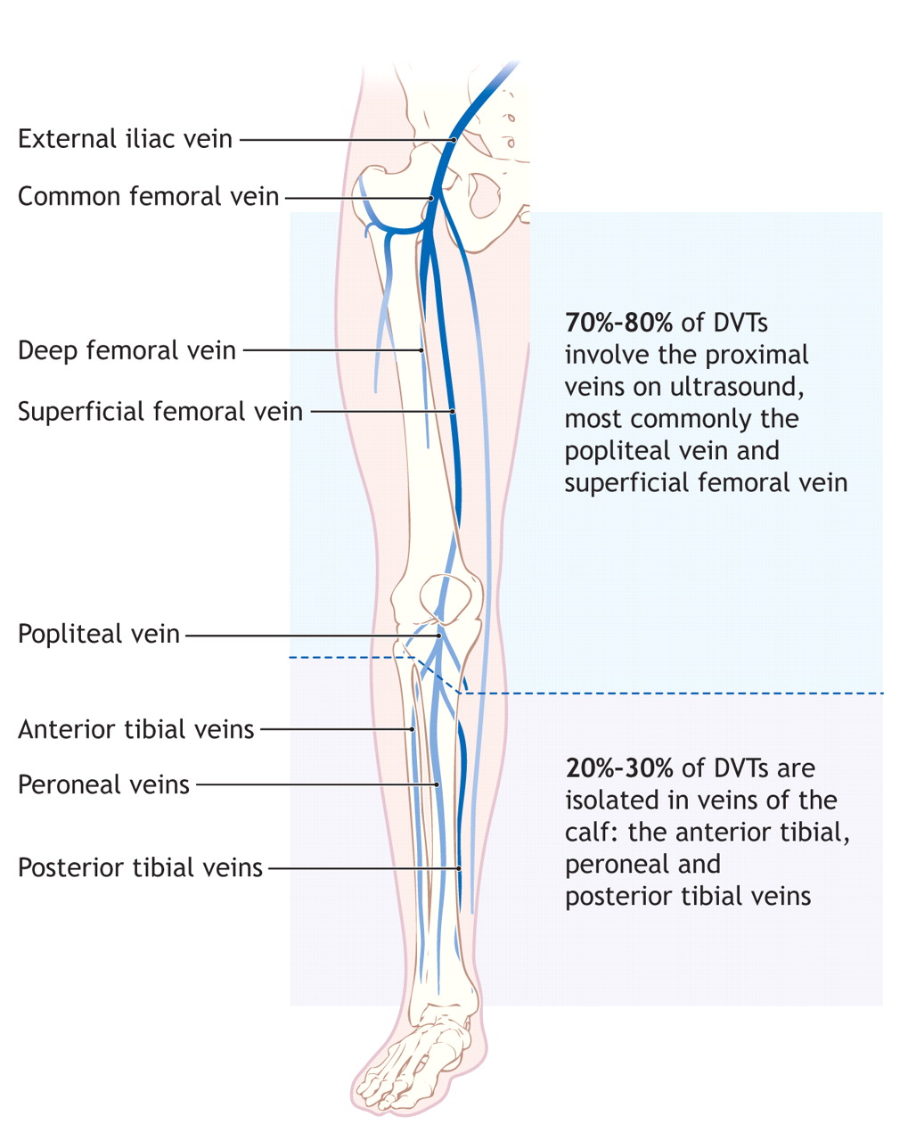
calf veins anatomy
Extremity Venous Anatomy and Technique for Ultrasound Examination we have 9 Images about Extremity Venous Anatomy and Technique for Ultrasound Examination like Anatomy Of The Lower Extremity Veins - Varicose Veins, Diagnosis and treatment of deep-vein thrombosis | CMAJ and also Extremity Venous Anatomy and Technique for Ultrasound Examination. Read more:
Extremity Venous Anatomy And Technique For Ultrasound Examination
 radiologykey.com
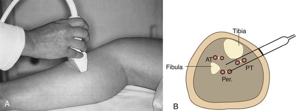
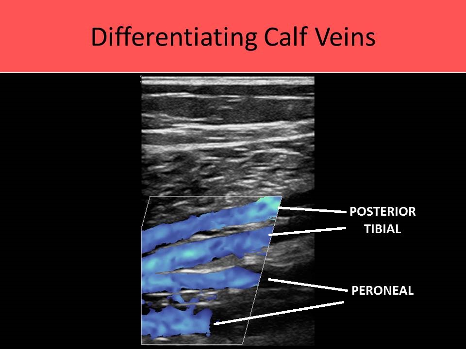
venous ultrasound extremity examination veins peroneal posterior tibial position technique anatomy duplex posteromedial viewing plane figure
Ultrasound Registry Review - Extremity Venous
 www.ultrasoundregistryreview.com
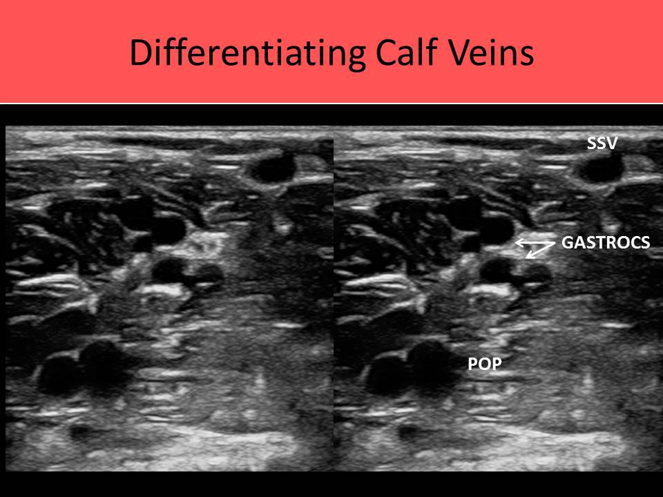
veins venous ultrasound soleal gastrocnemius anatomy extremity saccular calf dilated sinusoid sural
Calf Anatomy | All About The Calf Muscles
 www.kingofthegym.com
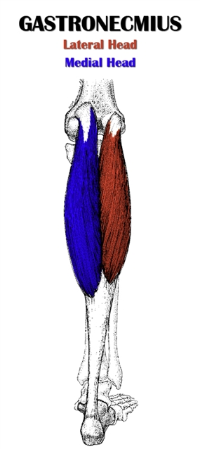
gastrocnemius anatomy calf muscle muscles gastroc soleus insertion medical action exercises characteristics include major following tendon kingofthegym
Anatomy Of The Lower Extremity Veins - Varicose Veins
 www.flandershealth.us
veins leg anatomy deep lower extremity varicose calf figure foot flandershealth
Ultrasound Registry Review - Extremity Venous
 ultrasoundregistryreview.com
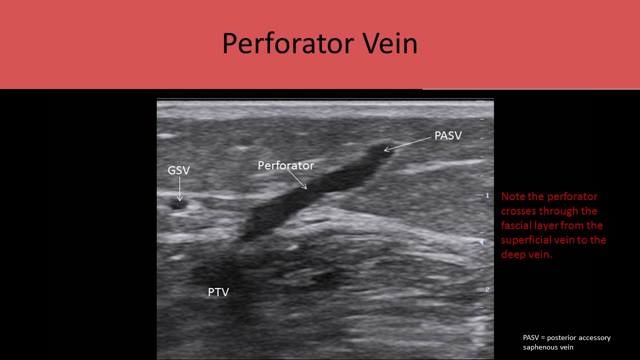
venous extremity ultrasound anatomy perforators
Ultrasound Registry Review - Extremity Venous
 ultrasoundregistryreview.com
ultrasound venous anatomy extremity ssv vascular vein gastrocnemius calf gsv muscle saphenous posterior sonography vessels registry
Diagnosis And Treatment Of Deep-vein Thrombosis | CMAJ
 www.cmaj.ca
vein thrombosis diagnosis cmaj 1087
Compartment Syndrome - WikEM
 www.wikem.org
wikem
DVT Ultrasound Protocol. Step By Step Guide To Ruling Out A DVT
 www.angiologist.com
dvt vein compression ultrasound duplex protocol rule step rules angiologist
Compartment syndrome. Veins venous ultrasound soleal gastrocnemius anatomy extremity saccular calf dilated sinusoid sural. Anatomy of the lower extremity veins