axial mri brain anatomy
Radiology - Normal brain anatomy - CT and MRI - YouTube we have 8 Images about Radiology - Normal brain anatomy - CT and MRI - YouTube like Axial view of co-registered normal CT and MRI scans - YouTube, MR-Eye: High-Resolution Microscopy Coil MRI for the Assessment of the and also Neurocysticercosis: nodular calcified stage | Image | Radiopaedia.org. Here you go:
Radiology - Normal Brain Anatomy - CT And MRI - YouTube
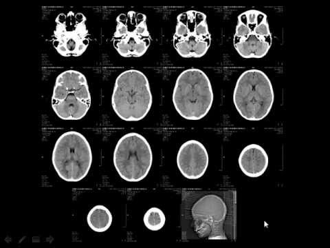 www.youtube.com
brain ct normal mri anatomy radiology
Axial View Of Co-registered Normal CT And MRI Scans - YouTube
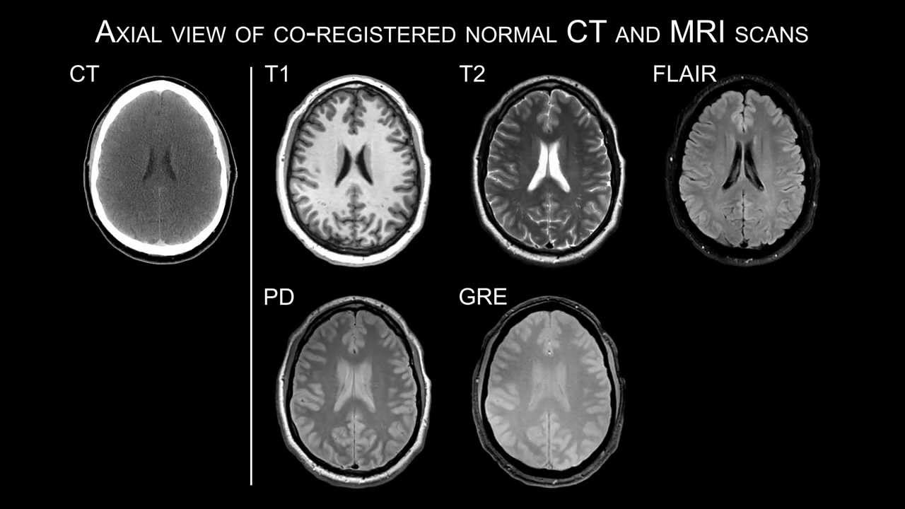 www.youtube.com
mri ct normal axial scans
Neurosurgical Procedures And Augmented Reality – Proof Of Concept
 futurebnd.com
reality augmented neurosurgical procedures proof concept holographic
Image | Radiopaedia.org
 radiopaedia.org
acoustic mri schwannoma neuroma intracanalicular vestibular nerve t2 vestibulocochlear radiopaedia wikidoc schwannomas tumours showing
MR-Eye: High-Resolution Microscopy Coil MRI For The Assessment Of The
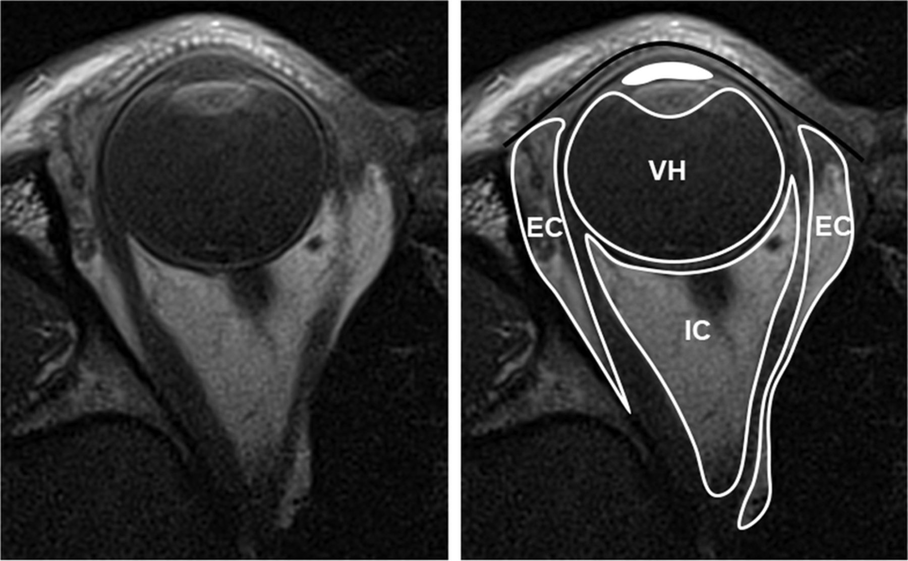 www.ajnr.org
mri ajnr oblique a6495
Insula: Aspect IRM
 info-radiologie.ch
insula irm axiale pondération sillon audiology radiologie
Neurocysticercosis: Nodular Calcified Stage | Image | Radiopaedia.org
 radiopaedia.org
neurocysticercosis calcified nodular stage radiopaedia case version
Radiological Anatomy: Calcarine Sulcus - Stepwards
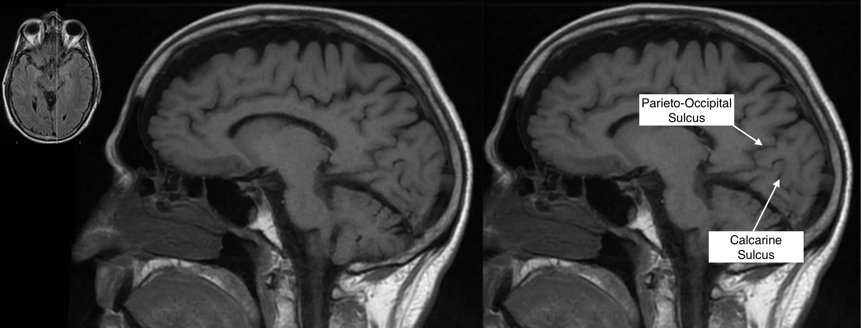 www.stepwards.com
calcarine sulcus stepwards radiological
Radiological anatomy: calcarine sulcus. Mr-eye: high-resolution microscopy coil mri for the assessment of the. Brain ct normal mri anatomy radiology